
Hip Dysplasia


What is Hip Dysplasia?
Hip dysplasia (HD) is abnormal joint laxity (looseness) of the hip joint. It is a common cause of hind limb lameness. This condition typically affects medium- to large-breed dogs. Many popular breeds commonly seen in the United States with this problem include Labrador Retrievers, Golden Retrievers and German Shepherds. Although more prevalent in these breeds, hip dysplasia can occur in dogs of any breed and size, including mixed breeds, and in cats. More than 125 dog breeds have been known to have a total hip replacement that was performed to correct hip dysplasia.

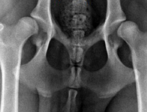
Normal Hips
The normal hip joint is a ball-and-socket joint. The “ball” (head of the femur) normally fits tightly in the “socket” (acetabulum) on the pelvis side of the joint. During growth and until adult skeletal structure is achieved, the bones, ligaments, muscles and other structures that cross the joint must develop at the same pace. When this occurs properly and in the presence of gravity, the joints develop normally without any laxity. The normal adult shape of a ball and socket are the result of all factors, including regular forces of the ball in the socket, developing normally.
Hip Dysplasia
With respect to hip dysplasia, the bones and soft tissues develop at a disproportionate pace, which results in bones that do not have a normal shape. The bones (both the ball and the socket) do not have a tight “fit” with one another because the bones do not have a normal shape, contributing to the joint laxity. Joint laxity stretches tissues that surround the joint, which leads to inflammation, pain, abnormal wear of the joint surfaces and pain manifested as lameness.
The cause of hip dysplasia is multifactorial. Genetics, diet, hormonal influence and environmental factors are all believed to contribute to the development of this condition. However, without abnormal DNA, the other factors likely have minimal or no influence. Extensive research is being conducted on the canine genome to identify the exact genetic code that is abnormal.
What are the symptoms of Hip Dysplasia?
Most dogs with HD show clinical symptoms by age one and some as early as six months. The symptoms are often subtle at first, with no obvious limp or possibly holding the leg up off the ground. Common observations include the following:
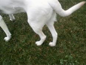 A “bunny hop” gait with both hind feet advancing at the same time instead of striding
- Hesitance to jump or go up a flight of stairs
- Premature tiring during exercise
- Content to “observe” rather than “participate” in vigorous play time
- Mild limp when rising after rest
- Weight shifting off of one or both hind feet while standing to eat or drink
- Slightly arched back as a result of weight shifting to the front legs
- Apathy toward exercise and play time
As with any joint problem, the body’s response is an attempt to stabilize laxity if it is present. When hip dysplasia is present, the degree of laxity varies from one dog to another, and there are times when one side is affected more than the other side. The laxity eventually results in joint inflammation known as secondary osteoarthritis (OA) at some point in life.
In secondary osteoarthritis, the inflamed tissues surrounding the joint thicken with scar tissue as part of the process in early arthritis. Some young dogs will actually improve as a result of Mother Nature’s course. Unfortunately, the “improvement” is usually temporary, with symptoms recurring. In almost every joint with HD that is abnormal at one year of age or less, the arthritic changes will progress. Dogs may be able to cope with the pain at certain stages, but eventually lameness or other symptoms will again be present. The time interval between subclinical symptoms is quite variable depending on the severity of the HD present initially.
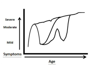 The severity of symptoms of hip dysplasia often wax and wane. The pain is sometimes severe at a young age and then improves only to return at some point. There are situations when the pain starts early in life (less than one year of age) and continues indefinitely unless treated. In other situations, the pain may be present, but the symptoms of lameness may not be obvious. Even with severe hip dysplasia, the limping is often only mild and easy to miss.
Dogs go through stages of coping with hip dysplasia pain, as the graph illustrates. Symptoms can vary throughout the dog’s lifetime but eventually are constant. Expert advice should be sought to determine appropriate treatment options.
How is the diagnosis of Hip Dysplasia made?
Radiographs (X-rays) are necessary to confirm the diagnosis of hip dysplasia. Before acquiring radiographs, the patient history and physical and orthopedic examinations are completed to determine if other causes of hind limb lameness are present.
What can be done if Hip Dysplasia is diagnosed?
Once the orthopedic exam and radiographs are completed, appropriate treatment options can be discussed. More than one option is almost always feasible. The surgeon and pet owner must consider the global perspective of each individual animal (age, breed, gender, body weight, body condition score, general health, intended function in life, presence or absence of concomitant problems, client constraints and personal decisions) to select the treatment which best fulfills all needs and expectations.
All options should be discussed along with the advantages and disadvantages while keeping the pet’s well-being in mind. The best outcome available is the restoration of successful and completely pain-free joints with normal biomechanical function. This means the dog will lead a healthy, happy normal life and be able to walk normally without pain or lameness.
Pain Management
Medical pain management may be a viable and effective treatment option for some dogs with mild hip dysplasia. This option is usually attempted when the diagnosis of mild hip dysplasia and/or osteoarthritis is first made.
Pain management only disguises the problem in dogs and cats with chronic, advanced, moderate, severe hip dysplasia and/or osteoarthritis in the hips. Costs accumulate rapidly with long-term use of pain and anti-inflammatory products for services that merely and temporarily mask symptoms. Prolonged symptomatic relief (more than one month) may just delay the inevitable while the arthritis continues to get worse and the dog suffers. This could potentially preclude a successful solution that was feasible earlier in life. Medical treatment and financial decisions should both be considered, especially if the dog or cat is young. The costs that accumulate during the animal’s lifetime could possibly be applied to a definitive and permanent surgical solution, such as a hip replacement.
A proactive, definite approach to resolve painful hips is a Total Hip Replacement (THR), which is the gold-standard surgical treatment for dogs and cats with moderate-to-severe hip dysplasia (HD). This means that it is the most effective, permanent and successful treatment available, allowing dogs (and cats) to resume a healthy, happy and normal life.
Total Hip Replacement Recovery Period
Dogs (and cats) recover much faster and better from hip replacement surgery than do humans. In fact, dogs (and cats) often do “too well, too soon” so owners must be diligent to “slow their animal companion down” during recovery. While all animals go through an anticipated recovery and healing period, they are typically walking on the new hip the day after surgery with only a minimal or mild limp.
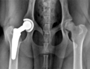
This is the appearance of a total hip replacement in a dog.
During the five to six weeks following surgery, activity should be limited. Dogs should walk on a good traction surface indoors and with a leash outdoors. No running, jumping, playing and going for long walks is allowed during this time. Regular walks typically begin about six weeks after surgery. The surgeon designs the exercise program. Activities progressively increase during the next couple months. The expectation is for dogs, including service dogs, hunting dogs, running companions, law enforcement and many other “dog occupations,” to resume normal activity when rehabilitation is complete.
Hip replacement surgery is not without some risk, just like in humans, but the risks are low. The risks should always be discussed and understood before surgery. Anesthesia is involved, which is as safe in animals as it is in humans. The risk of infection is low, but it is not zero-percent risk-free no matter what kind of surgical procedure is performed. Other risks often relate to the surgeon’s level of experience.
Femoral Head Ostectomy
It is possible to wait too long to opt for hip replacement surgery. Hip dysplasia can advance beyond the point where a hip replacement is no longer possible due to extensive bone being worn away along the acetabulum.

This is the appearance of the hip after a FHO.
If a total hip replacement is not feasible, a femoral head ostectomy (FHO) is a salvage procedure that can be performed. Unlike a hip replacement, biomechanical function is not maintained after the FHO. During this procedure, the ball component of the ball-and-socket hip joint is removed to create separation between the pelvis and the femur to decrease bone rubbing on bone. Rehabilitation is prolonged after a FHO and may take three to six months or more. Outcome depends in part of successful physical rehabilitation. Objective data on FHO patients shows that full recovery to normal function is considerably lower than after a hip replacement.
In conclusion, treatment of hip dysplasia should be carefully considered, and all options, including the advantages and disadvantages, should be discussed and clearly understood. The considerations should be discussed with your veterinarian prior to making any decisions.
William Liska, DVM, is a graduate of Iowa State College of Veterinary Medicine. Dr. Liska founded and now dedicates his career to Global Veterinary Specialists and the Global Veterinary Specialists Foundation.