Mast Cell Tumors (MCT) in Cats and Dogs

Mast Cell Tumors
Mast cell tumors are the most common skin cancer in dogs. While they also occur in cats, they are less common in that species. In addition to developing in the skin, they also can develop internally, especially in the spleen, which is seen more often in cats. Certain breeds are more likely to develop mast cell tumors, though any dog or cat can develop them. Dog breeds at higher risk for mast cell tumors include pugs, Boston terriers, boxers, Rhodesian ridgebacks, Shar-peis and Weimeraners. As far as cats go, the cutaneous form tends to be more common in the Siamese breed.
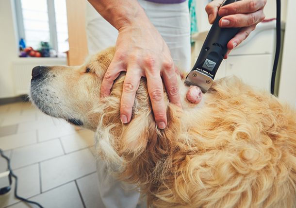
Mast cell tumors are known as the “Great Imposter” because they can masquerade as any other type of skin cancer. They can be flesh-colored or reddish, haired or hairless and may change in size (periodically enlarging or shrinking). The change in size comes from the inflammatory factors, including histamine, that are found in mast cell tumors. When the cells in the tumor become irritated, the inflammatory factors are released, leading to temporary swelling or enlargement of the tumor.
Diagnosis
Mast cell tumors of the skin can be diagnosed by taking a small sample of the area with a fine needle. Generally, your veterinarian can make the diagnosis based on this small biopsy during the appointment or within 24 hours. However, to determine the grade (severity) of the tumor, it will need to be fully removed and biopsied (examined by a veterinary pathologist). Since these tumors can have cells that invade nearby tissue, your veterinarian will take a substantial portion of the issue around the tumor when it is removed. Surgical removal also is the best treatment for your pet with this cancer.
Severity
After your veterinarian surgically removes the tumor, he or she will send it to a veterinary diagnostic lab where a pathologist will grade it to determine the level of the tumor’s severity. Grade I (low-grade) tumors are the least likely to have spread in the body, and surgery is considered curative for this grade of tumor. Grade III (high-grade) tumors are the most invasive and aggressive and the most likely to spread to other parts of the body. Grade II tumors are considered to be in between I and III in terms of invasiveness and aggressiveness.
Treatment
The type of treatment for your pet will depend on whether the mast cell tumor has spread in the body. At this point, your pet will undergo “staging” to determine if the cancer has spread to other areas, such as regional lymph nodes, distant lymph nodes or internal organs, such as the liver or spleen. Your veterinarian will perform lymph node aspirates, abdominal radiographs, an ultrasound and other diagnostics to ascertain how widespread the mast cells are in your pet.
Due to advances in veterinary medicine, tests can be applied to mast cell tumor biopsy samples to determine if certain chemotherapeutic agents should be used for treatment. There are several different chemotherapy protocols available. In some cases, the tumor may have been too large for your veterinarian to remove all of it, and the pathology report will indicate that the tumor has “dirty” margins, which means the pathologist is finding cancer cells at the edges of the biopsy sample. This most commonly happens when the tumor is very large or located on a limb or in the mouth, where it is difficult to make wide incisions around it and close the incision. If the tumor has not spread to other locations (localized disease), radiation therapy can be used to kill the remaining cancer cells at the surgical site. Most animals tolerate radiation therapy extremely well. If the tumor has spread to other areas or radiation is not an option, chemotherapy will be needed to cure the patient.
Identifying Severity of Mast Cell Tumors in Cats
Pathologists don’t grade feline mast cell tumors the same way they grade them in dogs. Some forms may generally be considered benign and surgery is curative. Other forms can be more invasive and aggressive and harder to completely remove. Further treatment, such as radiation or chemotherapy, may be recommended. The visceral mast cell tumors are the ones that are not located in the skin but found in the spleen, liver or gastrointestinal tract. If your cat is vomiting, has a decreased appetite and/or is losing weight, a mast cell tumor may be on your veterinarian’s differential diagnostic list. Surgery to remove the spleen may help in some of these cases, but in general, this form has a poor prognosis. As of now, no single chemotherapeutic protocol has shown to be very effective.
A Veterinarian’s Recommendation
If your pet is diagnosed with a mast cell tumor, have it biopsied and get a report from the pathologist. If surgery alone is not curative, ask your veterinarian about further treatment options or obtain a referral to a veterinary oncologist for continued care. Before starting treatments, such as radiation or chemotherapy, it is important to understand what your pet’s quality of life and potential life expectancy will be. These things are determined by the grade and stage of mast cell tumor your pet has, as well as other health issues that may be present. Your veterinarian or veterinary oncologist can help you determine the best choices for you and your pet.
Lori Teller, DVM is a graduate of Texas A&M College of Veterinary Medicine and lives in Houston, Texas. She practices at Meyerland Animal Clinic.

